Nati Salvadó,a* Salvador Butía and Trinitat Pradellb
aDpt. d’Enginyeria Química, EPSEVG, Universitat Politècnica de Catalunya, Av. Víctor Balaguer s/n, 08800 Vilanova i la Geltrú, Barcelona, Spain. E-mail: [email protected]
bDpt. Física i Enginyeria Nuclear, ESAB, Universitat Politècnica de Catalunya, Edifici ESAB, Campus del Baix Llobregat, Avda. del Canal Olímpic s/n, 08860 Castelldefels, Barcelona, Spain
Introduction
When viewing an art work, for instance a painting, we frequently have the wrong impression in that we think we are seeing the same as the artist painted and intended. We often forget that over the hundreds of years that may have elapsed, the painting has been subjected to an oxidising environment and in direct contact with many active substances. The materials used by the artist, although solid or semi-solid and thus not very reactive, are not fully inert and they have had a long time to react. One must also keep in mind the multiple invasive treatments and environments the artwork may have suffered over the years while being on display either in its original place or in private collections, on walls or stored in a museum.
The identification of materials and production techniques, aging processes, reaction compounds and impurities make the study of paintings an intriguing science from a chemical point of view, as well as the obvious historic and stylistic interest. Many of the substances we are interested in are found in very small proportions, deposited in sub-millimetre thick and very heterogeneous layers. Moreover, for obvious reasons, the amount of sample available for analysis is very small. All this makes their study a challenge for the scientist.
Microspectroscopy techniques play a very important role in taking on this challenge. Fourier transform infrared (FT-IR) microspectroscopy and Raman spectroscopy are both widely used for the study of these kinds of materials.1,2,3,4,5 Over the last few years the use of infrared radiation emanating from a synchrotron as a source for FT-IR spectroscopy has enabled better detection limits of certain substances and has opened new avenues for the research of ancient painting materials.1,2,3
Any information about the materials present in a painting is of great importance when proposing strategies of conservation and restoration for an art work. Moreover, it also offers the art historian new resources to establish relationships between painters and also to artist schools of the period.
The combination of using various analytical techniques on the same sample helps to overcome certain limitations and to obtain more precise information on the composition and distribution of the several substances present. Additional to the spectroscopy techniques herewith presented, X-ray diffraction with synchrotron radiation and scanning electron microscopy (SEM) are also commonly combined.
The change in the pictorial technique used in panel painting during the 15th century in Catalonia and, in general in the area under the crown of Aragon, make their study a subject of especial interest from the materials point of view.6 During this period, the egg yolk tempera traditional technique was substituted by a drying oil, i.e. linseed oil. However, both techniques are often found together in the same altarpiece. These two binders behave differently chemically. However, the reaction products are often the same since both contain lipids. The introduction of the oil technique in Catalonia points to the influence from the most active artistic areas of the period, in particular, from Flanders and Italy. The analysis of the painting materials is therefore of great interest to the art historians.
Case study
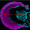
Bernat Martorell is one of the most representative Catalan painters of the first half of the 15th century. He used the egg tempera technique in all his paintings, at least in those studied up to now. Figure 1 shows a section of one painting sample extracted from a green area belonging to the clothes of a figure from “retaule de Sant Vicenç de Menàrguens” exhibited at the Museu Nacional d’Art de Catalunya (MNAC) in Barcelona. The green paint consists of a sequence of layers. The first layer is formed by a mixture of gypsum (CaSO4·2H2O) and binding media with a protein-based agglutinating substance that is related to animal glue. This layer was applied directly onto the wood surface, and acts as a ground layer over which the several chromatic layers are applied. This is the traditional technique used to prepare the wood surfaces in Catalonia and the introduction of the oil technique did not change this. The several painting layers are micrometres in thickness, but with adequate mechanical separation and thinning by means of a diamond-window compression cell, it is possible to obtain good transmission mid-IR spectra. Moreover, using a small aperture size (of about 10 μm × 10 μm) and the high signal-to-noise ratio achievable when synchrotron mid-IR radiation is utilised, adequate separation of the different compounds forming the layer may be obtained. This procedure allows for identifying most of the pigments, binders and also reaction and alteration compounds forming the layers.
, Photo: Calveras © MNAC-Museu Nacional d’Art de Catalunya. Barcelona. Bottom left: optical image of cross-section from green sample. Right: synchrotron-sourced mid-IR FT-IR microspectroscopy spectra corresponding to green sample from the altarpiece. Bottom: spectra from groundlayer. Middle, spectra from green layer. Top, spectra from varnish layers and altered surface layer. (Spectra were recorded on Beamline 11.1 SRS Daresbury Laboratory, UK, using a Nicolet Nexus FT-IR spectrophotometer, Nicolet Continuµm microscope, MCT detector, 128 scans, 4 cm–1 resolution, spot size 10 µm × 10 µm).")
In Figure 1 a sequence of mid-IR spectra recorded from the layers corresponding to the green paint (ground, chromatic layers and varnish) are shown. The green chromatic layer is formed by a mixture of green, white and yellow pigments mixed with egg yolk (appearing as a mixture of proteins and lipids). The green pigment identified corresponds to a mixture of copper-based compounds including copper hydroxychloride Cu2(OH)Cl3 (bands between 800 cm–1 and 1000 cm–1 are the planar deformation modes of the –OH group) and copper acetates. Copper-based green pigments such as, copper acetates, hydroxyacetates and hydroxychlorides obtained by the reaction of copper with vinegar vapours, were widely used during this period. Variations of the recipes used included the use of bronze and the addition of salt held together with honey or soap. This resulted in a large chemical variability of the compounds formed including several molecules associated with hydrated water and containing hydroxide groups, which makes the study of these pigments particularly complex and interesting. These compounds usually show a turquoise hue, which was corrected by mixing them with yellow and white pigments. The white pigment is lead white; a mixture of lead basic carbonate, hydrocerussite 2PbCO3·Pb(OH)2, and lead carbonate, cerussite PbCO3. Lead white was the most commonly used pigment for centuries. The yellow pigment identified is a lead stannate widely used in the period, which has its origin in the glass industry.
The pigment used by Bernat Martorell is the lead stannate of type II, Pb(Si,Sn)O3 which differs from type I by the presence of silicon substituting tin and by its crystallographic structure. The use of this pigment corroborates his Italian influence contrasting with other contemporary painters closer to the Flemish school. In the mid-IR region the lead tin yellow type II pigment is identified by the Si–O stretching vibration bands, while the type I pigment is transparent for wavenumbers above 60 cm–1. Consequently, it is possible to differentiate type I and type II pigments in the low frequency region <600 cm–1. The use of synchrotron sourced far-IR radiation with FT-IR microspectroscopy coupled with a bolometer detector allows studies in the far-IR region and thus expands the possibilities of this technique in the study of ancient paintings. However, diffraction effects require the use of a bigger spot size (of about 50 µm diameter) than for the mid-IR region, thus limiting the separation of compounds that may be obtained. Figure 2 shows both the mid- and far-IR regions corresponding to three yellow paints from the period studied; the top spectrum corresponds to the same sample shown in Figure 1, the middle and bottom spectra correspond to a lead stannate of type I mixed with egg yolk and drying oil, respectively. Both compounds show characteristic bands in the far-IR region. Both yellow pigments may also be identified using Raman microspectroscopy (characteristics bands at 78 cm–1, 128 cm–1, 196 cm–1, type I, and 69 cm–1, 139 cm–1, type II); Figure 3 shows the Raman spectra corresponding to the three same painting mixtures as shown in Figure 2.
. Middle: from green layer from the sample of the altarpiece of “Conestable” by Jaume Huguet (15th century, Santa Àgata chapel, Barcelona). Bottom: from green layer from the sample of “Sant Vicenç de Sarrià” by Bernat Martorell (15th century, MNAC). (Spectra recorded using Beamline IR 1 on the synchrotron light source ANKA, Germany, and a Bruker IFS 66v/s, Bruker IRScope II microscope, MCT mid-IR detector and 4.2 K bolometer for the range 700–200 cm–1, 128 scans, 4 cm–1 resolution, spot size 10 µm × 10 µm and 50 µm × 50 µm for the mid-IR and far-IR regions, respectively).")
.")
On the top of the chromatic layers, a varnish is often applied to give brilliancy and protection to the paint. Obviously, those are the layers appearing more altered and which have undergone more interventions and atmospheric depositions. Although there is no direct evidence of their use in the period studied, varnishes were often applied later to protect the paintings. Figure 1 shows the presence of two different vanishes. The inner layer corresponds to a triterpenoid resin, while the most external layer corresponds to a modern acrylic substance that was applied in the latest restoration of the artwork. Although varnishes are known to have been used since very ancient times, it is not until the 16th century that they were regularly used in paintings to protect the chromatic layers. Triterpenoid resins have been used for centuries and therefore it is not possible to know to which historic period they correspond.
Finally, the IR spectra corresponding to the chromatic layers show also the presence of some carboxylate salts, compounds formed by the reaction of the free fatty acids, produced by aging of the lipid substances, with metal ions such as lead and copper from the pigments. However, previous studies have shown a preference for copper if present in the formation of carboxylates. In the case studied here copper carboxylates have been formed. Oxalates are also commonly found either in the chromatic, ground or varnish layers. Their origin may be related to the degradation of the organic matter which sometimes has been associated to the bioactivity.
Summary
The combination of different techniques is indispensable for a good characterisation of the materials forming ancient paintings, and in particular when the identity of compounds present in minor amounts are to be determined. The use of synchrotron radiation as a light source provides advantages, since it produces better quality spectra (better signal-to-noise ratio) and a better spatial separation of compounds due to the small spot size used.
Acknowledgements
This work was funded by the EU IA-SFS MS-97 project at ANKA (far-IR microspectroscopy measurements), the 45256 project at SRS (beamline 11.1) Daresbury Laboratory (mid-IR microspectroscopy measurements), and 20090887 project (beamline SIMS) at Soleil Paris (Raman spectroscopy measurements). N. Salvadó and S. Butí received financial support from the “Ministerio de Ciencia e Innovación” (Spain) Grant HAR2009–10790.
Part of this work was carried out within the framework of agreements of collaboration between the Technical University of Catalonia (UPC) and the “Museu Nacional d’Art de Catalunya” (MNAC).
References
- N. Salvadó, S. Butí, M.J. Tobin, E. Pantos, J.N.W. Prag and T. Pradell, “Advantages of the use of SR-FTIR microspectroscopy: Applications to Cultural Heritage”, Anal. Chem. 77, 3444 (2005).https://doi.org/10.1021/ac050126k
- N. Salvadó, S. Butí, J. Nicholson, H. Emerich, A. Labrador and T. Pradell, “Identification of reaction compounds in micrometric layers from gothic paintings using combined SR-XRD and SR-FTIR”, Talanta 79, 419–428 (2009).https://doi.org/10.1016/j.talanta.2009.04.005
- M. Cotte, P. Dumas, Y. Taniguchi, E. Checroun, P. Walter and J. Susini, “Recent applications and current trends in Cultural Heritage Science using synchrotron-based Fourier transform infrared micro-spectroscopy”, CR Phys. 10, 590–600 (2009).https://doi.org/10.1016/j.crhy.2009.03.016
- S. Prati, E. Joseph, G. Sciutto and R. Mazzeo, “New advances in the application of FTIR microscopy and spectroscopy for the characterization of artistic materials”, Accounts Chem. Res. 43, 792–801 (2010).https://doi.org/10.1021/ar900274f
- R.J.H. Clark, “Pigment identification by spectroscopic means: an arts/science interface”, CR Chim. 5, 7–20 (2002).https://doi.org/10.1016/S1631-0748(02)01341-3
- N. Salvadó, S. Butí, F. Ruiz-Quesada, H. Emerich, T. Pradell, Mare de Déu dels Consellers and de Lluís Dalmau, “Una nova tècnica per a una obra singular”, Butlletí del MNAC 9, (2008).