Articles and Columns
The Foundation for Analytical Science & Technology in Africa (FASTA) is a charitable company that was established in 2005 in response to a request to provide a GC-MS to the Jomo Kenyatta University of Agriculture & Technology (JKUAT) in Nairobi, Kenya. FASTA was founded by Steve Lancaster of BP and Barrie Nixon of Mass Spec UK Ltd. The objectives of the organisation are to support scientific education, analytical research and the preservation of the environment in Africa via capacity-building and technology transfer.
After the progressive development of techniques that yielded results for optimal samples, Laser Ablation Inductively Coupled Plasma Mass Spectrometry (LA-ICP-MS) has at last provided a means of analysing individual fluid inclusions in typical, rather than exceptional, samples. The first truly quantitative LA-ICP-MS analyses of single fluid inclusions were carried out in ETH Zurich in a group led by Christoph Heinrich and Detlef Gunther. They addressed the key issues of ablating into transparent host crystals to release fluid in a controlled manner, minimising interferences and finding suitable calibration strategies, while at the same time quantifying a signal that is typically released over a time of just a few seconds, giving a brief surge in the signal but no definite plateau. The laboratory we have built up in Leeds is based on theirs, but we have significantly developed the data handling.
Christopher Burgess
Burgess Analytical Consultancy Limited, “Rose Rae”, The Lendings, Startforth, Barnard Castle, Co. Durham, DL 12 9AB, UK
John Hammond
Starna Scientific Ltd, 52–54 Fowler Road, Hainault Business Park, Hainault, Essex, IG6 3UT, UK/p>
In the present study, operando infrared (IR) spectroscopy was used to investigate under realistic conditions the oxidation activity of Pt and Pd supported on different oxides, with the aim of generating mechanistic information that will be used for the design of improved formulations.
Tom Scherzer, Gabriele Mirschel and Katja Heymann
Leibniz Institute of Surface Modification (IOM), Permoserstr. 15, D-04318 Leipzig, Germany. E-mail: [email protected]
Scientific studies of artworks are an important practice in many institutions dedicated to the study and protection of cultural heritage. Applied physics and chemistry provide the scientific data necessary to characterise and understand the origin, the degradation processes and the environment in which the artwork was created or has existed.
Peter J. Jenks
the Jenks Partnership, Newhaven House, Junction Road, Alderbury, Salisbury, Wiltshire SP5 3AZ, UK. E-mail: [email protected]
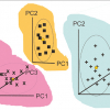
In our previous column we introduced CVA, one of the very early applications of multivariate analysis (1930s). In this column we will discuss SIMCA (officially it is Soft Independent Modelling of Class Analogies, but no one uses the long form!). SIMCA was invented 30 years later by another pioneer, Svante Wold (the man who coined the word “chemometrics”).
Maria C. Prieto Conaway,a Shousong Cao,b Farukh Durrani,b Youcef Rustum,b Ping Wang,c Khin Marlarc and Latif Kazimc
aThermo Fisher Scientific, San Jose, CA, USA
bRoswell Park Cancer Institute, Department of Cancer Biology, Buffalo, NY, USA
cRoswell Park Cancer Institute, Department of Cell Stress Biology, Buffalo, NY, USA
As the column title rightly suggests Quality does Matter. The authors look at the major changes that have taken place over the last 50 years in four key areas: The National Metrology Institutes (NMIs), the instrument manufacturers, the user base and the globalisation of regulation through international regulatory bodies.
Our laboratory has received several requests from public or private institutions to solve problems related to conservation and restoration of samples from cultural heritage. A brief description of the FT-IR methodologies used to solve some of them are detailed within this article.
Antony Neil Daviesa and Jörg Ingo Baumbachb
aExternal Professor, University of Glamorgan, UK. Director, ALIS Ltd, and ALIS GmbH–Analytical Laboratory Informatics Solutions
bISAS–Institute for Analytical Sciences, Metabolomics Department, Bunsen-Kirchhoff-Str. 11, 44139 Dortmund, Germany
Inductively coupled plasma-mass spectrometry (ICP-MS) was introduced commercially in 1983 as a very sensitive analytical technique to be deployed for (ultra)trace element analysis. Compared to the previously existing techniques of atomic absorption spectrometry (AAS) and ICP-optical emission spectrometry (ICP-OES), the main advantages offered by ICP-MS over these techniques were its pronounced multi-element capabilities and substantially higher detection power, respectively.
L. Rello,a E. García-Ruiz,b M.A. Belarrab and M. Resanob*
aDepartment of Clinical Biochemistry, “Miguel Servet” Universitary Hospital, Paseo Isabel La Católica 1–3, 50009 Zaragoza, Spain
bDepartment of Analytical Chemistry, University of Zaragoza, Pedro Cerbuna 12, 50009 Zaragoza, Spain. E-mail: [email protected]
During the last few decades, solution and solid state techniques have been utilised to obtain information about the properties of supramolecular host–guest complexes. Mass spectrometric analysis of these fragile non-covalent complexes has been focused on the determination of the molecular mass of the interacting molecules and the analysis has concentrated on the characterisation of covalent compounds. Since the invention of the soft ionisation techniques [namely ESI (electospray ionisation) and MALDI (matrix-assisted laser desorption/ionisation)] and their development for mass spectrometry (MS) instruments, the area and way that MS analysis is used have greatly changed and expanded. In particular, ESI has attained a steady position for the analysis of biomolecules, their non-covalent complexes and other rather fragile systems, which were earlier impossible to study by mass spectrometric methods. Today, MS can be employed not only for molecular weight identification purposes but also for sophisticated analyses on versatile properties of compounds. In the area of supramolecular chemistry, MS studies are becoming more and more general, although MS utilisation is still quite limited.
A.M.C. Daviesa and Tom Fearnb
aNorwich Near Infrared Consultancy, 75 Intwood Road, Cringleford, Norwich NR4 6AA, UK. E-mail: [email protected]
bDepartment of Statistical Science, University College London, Gower Street, London WC1E 6BT, UK. E-mail: [email protected]
Patrick Garidel
Boehringer Ingelheim Pharma GmbH & Co. KG, Process Science, Pharmaceutical Basic Development, D-88397 Biberach an der Riss, Germany. E-mail: [email protected]
Peter J. Jenks
the Jenks Partnership, Newhaven House, Junction Road, Alderbury, Salisbury, Wiltshire SP5 3AZ, UK
During the last decade, the amount of counterfeit drugs on the worldwide market has rapidly increased. It is difficult to determine the exact scale of this problem, since not all counterfeit drugs are reported or even detected. The drugs that are often counterfeited can be divided into two groups, depending on the region where they are brought onto the market. The use of Raman spectroscopy in the pharmaceutical field has been increasing over the last few years. Raman spectroscopy has some specific benefits, which can be very useful for the detection of counterfeit drugs: a Raman spectrum can be recorded rapidly without any sample preparation. Moreover, not only does a Raman spectrum provide information about the active ingredient present in the drug, but also a Raman spectrum contains information on its concentration and allows identification of excipients present in the drug. So far, this technique has been used for the detection of different kinds of illicit drugs, such as cocaine, heroin and ecstasy, and also for the detection of counterfeit antimalarial and Viagra® tablets. This article will demonstrate the use of Raman spectroscopy as a fast and easy detection system for different counterfeit erectile dysfunction drugs. Three different genuine erectile dysfunction drugs are available on the market: Viagra®, Cialis® and Levitra®. Viagra® is the most commonly counterfeited drug, but the amount of counterfeit Cialis® has rapidly increased over the last few years. The possible counterfeits analysed consisted of two Viagra® tablets, SEYAGRA-GEL containing the same active ingredient as Viagra® and two tablets claiming to contain the same active ingredient as Cialis®.
The ultimate use of XRF for medical analysis is in vivo measurements made directly in the living patient or volunteer. It started with quantitative analysis of iodine in the human thyroid. The idea sprang from the pioneering work by Jacobsson, who developed a technique for subtraction radiology of iodine using two x-ray energies, one above and one below the K-absorption edge of iodine. Hoffer et al. realised that if that technology worked, there should be a chance to see the emitted characteristic x-rays from iodine using the semiconductor detectors, which at that time had been developed for nuclear and particle physics. In this way the first in vivo XRF analysis was done, quantifying the iodine concentration in human thyroid, typically around 400 µg g–1. The further development of the in vivo XRF technique was related to the analysis of heavy elements, first covering lead and later cadmium and to some extent also mercury in occupationally exposed workers. Platinum was also analysed to investigate uptake and kinetics of the cytostatic agent cis-platinum in tumour patients. The following section describes efforts made to study various toxic elements in vivo in occupationally exposed workers and in patients.


