Natalie Z.M. Homer
CRF Mass Spectrometry Core, Centre for Cardiovascular Science, Queen’s Medical Research Institute, University of Edinburgh, Edinburgh EH16 4TJ, UK. E-mail: [email protected]
Introduction
The application of mass spectrometry (MS) to clinical analysis has evolved considerably since it was first used, both in research facilities and for routine analysis in clinical biochemistry laboratories. Mass spectrometry is a technique that measures ions in the gaseous state. Samples are introduced into an ion source, ionised and then separated in a mass analyser according to their mass-to-charge ratio (m/z). Coupled to chromatographic separation techniques such as gas chromatography (GC) or liquid chromatography (LC), mass spectrometry has long been recognised as the gold standard for validation of quantitative analytical assays.
For successful MS analysis the correct analytical procedure, i.e. extraction and sample preparation and the best instrumentation, in terms of chromatography and mass spectrometer set up, must be chosen. Initially GC-MS was used for biological analysis but the requirement that GC needs a volatile analyte meant that elaborate extraction and derivatisation protocols were needed for analysis of the typically polar, thermally labile and involatile biomolecules found in clinical samples. LC is a much more appropriate separation technique for these polar, thermally labile molecules found in biological samples. It also reduces the need for derivatisation to volatile molecules. It is the coupling of LC and MS that led to the adoption of clinical MS. The very recent popularity of clinical MS can in part be attributed to the unique ability of MS to detect multi-analytes with high specificity.
Sample preparation and chromatography
Clinical samples are complex biological matrices and contain interferences that can lead to so-called matrix effects within the mass spectrometer. Samples are usually prepared for quantitative MS analysis by addition of an internal standard followed by extraction to remove as much of the interferences as possible. Extraction methods commonly used for clinical samples include liquid–liquid extraction (LLE), solid-phase extraction (SPE) and simple protein precipitation with a solvent. If sample clean-up is not sufficient it can lead to matrix effects, including ion suppression of the analyte, usually observed as a loss of response. This negatively affects the detection limits, accuracy and precision of the assay. Various ion suppression tests have been developed and these are an important part of the method validation set up required for all clinical MS assays. The two most effective ways of avoiding ion suppression are improved sample extraction and optimised chromatographic selectivity.
Once prepared a sample is introduced into a chromatography system, which consists of a pump and an analytical column. The purpose of the chromatography system is to separate the components of the sample as much as possible prior to introduction into the MS. The analytical column is usually reversed phase, which means that the silica bead packing stationary phase is chemically modified and more aqueous solvents are used as the mobile phase. The chemical composition of the stationary phase dictates the selectivity of the column, i.e. order of elution and peak shape.
Analytical LC column technology is continuously improving. The better the resolution, which is simply how well separated each peak is, the better the assay. Sub-2 µm particles have been introduced; these generate sharp peaks and excellent resolution with improved capacity. However, the small particle size leads to high backpressure and requires LC pumps that can withstand these ultra higher pressures (uHPLC, ultra high performance LC). In response to this, LC columns packed with fused-core particles >2.5 µm have been developed to allow separation comparable to sub-2 µm particles but without the requirement of new LC pumps that need to withstand high pressures.
MS instrumentation
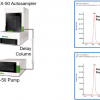
An overview of how a typical chromatograph-mass spectrometer is set up is shown in Figure 1. The ion source and mass analyser of the MS have had continuing research and development over the past three decades and these advances have led to the different types of MS used in the clinical setting today.

Following LC separation by the chromatography system the sample is introduced into an ion source at the front end of the MS. Ionisation modes include electron ionisation (EI), chemical ionisation (CI), atmospheric pressure ionisation (API), which incorporates electrospray ionisation (ESI) and atmospheric pressure chemical ionisation (APCI), and finally matrix-assisted laser desorption/ionisation (MALDI). ESI is most typically used to analyse the biomolecules encountered in clinical samples.
Once ionised the mass analyser separates the ions according to their m/z. Mass analysers include magnetic or electric sectors, time-of-flight (ToF) instruments, quadrupoles and two-dimensional and three-dimensional ion traps. The magnetic sector mass analyser is the classic apparatus for mass separation. It is this type of high resolution MS instrument that is required for analysis of dioxins. However, softer ionisation techniques which lose specificity such as ESI, have led to the use of quadrupole mass analysers, where a quadrupole consists of four parallel rods or poles, generally of hyperbolic cross-section, through which ions are passed and separated (Figure 2).

The introduction of tandem mass spectrometry (MS/MS) increased the specificity of MS significantly. This is when two or more mass analysers are placed in sequence and fragmentation of the ions is induced in a collision cell to give structural information. Trace analysis of complex biological matrices is ideally suited to tandem MS instruments. The most commonly encountered MS/MS is a triple quadrupole, which has a mass analysing quadrupole, a quadrupole collision cell and a third mass analysing quadrupole. The greater sensitivity in conjunction with the improved specificity of MS/MS over immunoassays has led to it becoming the preferred mode of analysis, particularly in the field of clinical endocrinology. In 2007 the American Endocrine Society, recognising the importance of MS/MS, issued a statement recommending the use of LC-MS/MS for the determination of endogenous steroid hormones over more traditional technologies such as immunoassays.1
Examples of clinical applications of MS
Much of the uptake of MS in the field of clinical research has been driven by the technological advances within chromatography and MS. Brought together, the advantages of LC, API and MS/MS have been exploited and a number of assays for clinical diagnosis are now routinely used. The analysis of steroid hormones by MS is a well-documented area of research. Other commonly encountered uses include newborn screening for congenital metabolic diseases such as aminoacidopathies and fatty acid oxidation disorders, multi-analyte therapeutic drug monitoring, oncology drugs, anti-virals, toxicant and drugs of abuse screening and analysis of endogenous peptides.2
In recent years there has been much emphasis placed on fast chromatographic analysis. The push for fast analysis in the clinical laboratory has become possible with the introduction of uHPLC and short analytical columns, enabling exceptionally short run times, of less than 5 min. This high throughput is desirable when faced with large sample numbers and enables quick return of data. However, there is a lot of discussion as to whether the quality of data is compromised. Protein precipitation as a mode of sample clean-up should certainly be avoided in fast analyses as matrix ion suppression is a problem in this kind of sample. If samples are not well extracted and run times are so short that matrix effects become a problem then the quality of data can be affected. A further concern is the co-elution of biologically similar compounds, as in the fast analysis of androgens. Testosterone (T) has a biologically inactive isomer, epi-testosterone (epiT). These isomers behave identically in the MS as they are isobaric, but can be resolved chromatographically given the correct conditions. It is very important that they are resolved as otherwise the biologically inactive epiT may contribute to the signal of T resulting in a false result. Many fast analyses do not allow complete resolution of these isomers and caution should be taken when accepting and interpreting results obtained in this way. In this situation a longer analysis time with complete isomer resolution should be chosen, instead of fast analysis compromising the data quality.
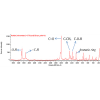
Advances in sample handling and improvements in chromatography instrumentation have allowed the development of some exciting clinical applications. On-line multi-dimensional chromatography technology allows an unextracted sample to be introduced into the chromatography apparatus and can also lead to faster analysis. These systems generally consist of multi-channel switching valves, on-line SPE cartridges and analytical columns ahead of ESI-LC-MS/MS. Steroids and isoprostanes are often analysed in this manner. In these assays it is important to eliminate ion-suppresssing interferences where possible and on-line SPE has been succesful in achieving this. A recent assay established to analyse thyroxine3 used dialysis and on-line SPE for an ESI-LC-MS/MS assay for their fast determination by injecting onto a C5 guard column, washing and eluting onto a C18 analytical column. A steep gradient eluted analytes in less than 3 min and all quantitative criteria were met using this method.
The use of steroid panels is of particular interest for the diagnosis and treatment of complex endocrine disorders such as primary hyperaldosteronism, adrenal insufficiency, congenital adrenal hyperplasia (CAH), Cushing’s syndrome and gonadal dysfunction. Multi-steroid analysis is a challenging diagnostic clinical requirement that benefits greatly from multi-dimensional LC-MS/MS. Thirteen different steroids have been successfully determined in protein-precipitated serum using a 2-D system in which a POROS® column is used for on-line SPE and a monolithic C18 analytical column is used to rapidly separate in a total run time of 4 min by ESI-LC/MS/MS with a linear ion trap.4
In addition to using on-line multi-dimensional chromatography, it is possible to allow multiplexed sample introduction whereby up to four channels can be fed into one MS/MS. This means that the MS is being used highly efficiently as it is not dedicated to a long run, waiting for the important 1–2 min time window of elution of the analytes. Instead, the on-line extraction is staggered and the mass spectrometer receives the expected analyte eluate time-window in succession.
Taking advantage of MALDI ionisation, blood spots, which have traditionally been reserved for simpler analytical techniques, can also be analysed by MS. This is an unusual combination of MALDI-style ionisation and LC-MS. An example of this technique being used clinically is through newborn screening for phenylketonuria (PKU), which is a congenital disorder that renders phenylalanine hydroxylase dysfunctional. This enzyme is responsible for the conversion of phenylalanine (Phe) to tyrosine (Tyr). By measuring both the substrate Phe and the product Tyr from a newborn bloodspot by MS/MS and determining the ratio of concentrations it is possible to detect PKU in the newborn with a very high sensitivity and specificity, higher than measuring Phe alone which is made possible by MS/MS.5
Having exploited API and MALDI ionisation, triple quadrupole technology and the advancements in LC and analytical columns, clinical mass spectrometry continues to be at the forefront of applied MS technology. However, one area that is not fully exploited yet is that of high resolution MS (HRMS). The accurate mass determination afforded by HRMS allows monitoring over a defined mass range, which differs from the targeted analysis approach used by triple quadrupole analysis. In targeted analysis there is no chance of detecting non-target analysis. A recent review has suggested that the advantages of high-resolution mass analysers based on ToF and Fourier transform instruments, which have a similar cost to triple quadrupole instruments, should be used in clinical MS. This is in contrast to FT-ICR (Fourier transform ion cyclotron) MS and sector instruments which have very high purchase and maintenance costs. The accurate mass determination of HRMS is well suited to screening applications.6 Further improvement is needed in the technology, however, to improve the quantitation in high-resolution instruments due to the limited linear range compared to triple quads. At this time HRMS is mostly restricted to research laboratories.
Summary
The range of clinical applications outlined briefly here is broad and constantly expanding. Applications include analysis of small molecules, multi-steroid panels, newborn screening and targeted proteins. In addition, much research is being conducted in the pioneering fields of proteomics and metabolomics. The diversification and improvements in LC and MS technology seen over the past few decades including the use of tandem mass spectrometry, different mass analysers, improved ionisation sources, the introduction of ultra high pressure LC and on-line extraction and multiplexing mean that questions are being answered in clinical MS laboratories that a decade ago were only attempted in research laboratories. There is no doubt that MS will continue to feature heavily in the clinical biochemistry laboratory and with the continued technological improvements MS will certainly function as an important clinical research tool.
References
- W. Rosner, R.J. Auchus, R. Azziz, P.M. Sluss and H. Raff, J. Clin. Endocrinol. Metab. 92, 405–413 (2007).
- B. Shushan, Mass Spectrom. Rev. 29, 930–944 (2010).
- B. Yue, A.L. Rockwood, T. Sandrock, S.L. La’ulu, M.M. Kushnir and M.W. Meikle, Clin. Chem. 54, 642–651 (2008).
- U. Ceglarek, L. Kortz, A. Leichtle, G.M. Fiedler, J. Kratzsch and J. Thiery, Clin. Chem. Acta 401, 114–118 (2009).
- D.H. Chace, T.A. Kalas and E.W. Naylor, Clin. Chem. 49, 1797–1817 (2003).
- J.-L.H. Jiwan, P. Wallermacq and M.-F. Hérent, Clin. Biochem. 44, 136–147 (2011).