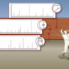
At any given moment, a variety of dynamic processes occur inside a cell, with many developing over time. Because current research methods for gene profiling or protein analysis destroy the cell, study is confined to just that one moment in time, and researchers are unable to return to the cell to examine how things change beyond that snapshot. A team led by Northwestern Engineering scientists has developed a minimally invasive method to sample cells that can be repeated multiple times, one of the first to do so. The process, called localised electroporation, has implications in studying processes that evolve, such as cells’ response to treatments for cancer and other diseases.
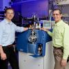
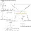
Horacio Espinosa led the team that created the live cell analysis device (LCAD), which can non-destructively sample the contents from small number of cells many times. When LCAD is coupled with self-assembled monolayer desorption ionisation (SAMDI), a highly sensitive and label-free method for quantification of enzymatic activity using mass spectrometry, the intracellular contents sampled by the LCAD are then analysed for the presence of enzymes. SAMDI was developed in the lab of Milan Mrksich at Northwestern University.
“By exploiting advances in microfluidics and nanotechnology, localised electroporation can be employed to temporarily open small pores in the cell membrane enabling the transport of molecules into the cells or extraction of intracellular contents. Since the method is minimally invasive to the cells, it can be repeated multiple times without their disruption”, Espinosa said.
“Certain enzymes may be linked to disease pathways, such as certain types of cancers, and they may be the target of therapeutics. Using this platform, it is now possible to study how enzymatic activity varies between healthy cells and cells from a tumour biopsy”, Mrksich said.
The LCAD-SAMDI platform offers an opportunity for biologists and physicians to investigate how specific treatments may alter these enzymatic activities and the associated diseases over time.
“The platform is one of the world’s first technologies allowing this type of research, a biopsy but performed on cells at the nanoscale”, Espinosa said.
John A. Kessler study co-author added “without disrupting the cell, it provides a window to processes inside cells and enables research that can determine the quantity of an active enzyme, how enzymatic activity in cells changes over time, and what changes in the activity occur in response to a treatment.”
This method opens up the possibility to investigate time-dependent processes, like cell differentiation, disease progression or drug response, at regular intervals.
“We envision that this technique can be used in scenarios such as screening drugs or designing and optimising treatment courses that can arrest disease progression in cells”, Espinosa said.
Most established methods require killing the cells being analysed. Currently, complex computational methods are used for retrieving temporal information from single snapshots, but assumptions about the dynamics and limitations on the time scales and scenarios remain. The LCAD also can be used to deliver proteins into cells. The combination of delivery and sampling could potentially be used in studies involving delivery of molecules, like DNA and proteins, and investigating its effect on the activity of another via sampling.
“We have used the same concept of localised electroporation to do CRISPR gene editing and we are now using machine learning to automate the process”, Espinosa said.
Overall, this method can provide complementary information regarding cellular dynamics, which may not be possible using traditional assays. In the future, as the technology improves and sensitivity increases, it may be possible to sample temporal information for several different types of proteins simultaneously from the same cell populations.
The research was published in Small.


