
: lipids (red), proteins (green) and carbohydrates (blue). © Helmholtz Zentrum München/Miguel A. Pleitez")
Metabolic diseases such as diabetes and obesity are ever more common globally. In addition to genetic disposition, lifestyle contributes strongly to their prevalence. Precise monitoring methods are needed in order to, for example, evaluate how a change in diet or exercise affects disease and its metabolic characteristics. A team from the Institute of Biological and Medical Imaging at Helmholtz Zentrum München and at the Chair of Biological Imaging at TranslaTUM at the Technical University of Munich has developed a technique that provides real-time images of biomolecules in living cells without the need for labels or contrast agents. The evaluation of the imaging system was performed in collaboration with the Institute for Diabetes and Cancer at Helmholtz Zentrum München and the Heidelberg University Hospital.
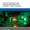
The new technology is based on photoacoustic spectroscopy and is called Mid-infraRed Optoacoustic Microscopy (MiROM). Specific molecular vibrations are targeted with mid-infrared lasers, triggering a thermoelastic expansion, the ultrasound waves from which are detected and processed to form an image of the distribution of specific molecules, depending on the wavelength(s) of excitation.
Compared to previous techniques, one significant advantage of this new method is that it is no longer limited to dry-fixed samples. By detecting acoustic waves, which pass easily through tissue, rather than photons that are strongly absorbed by tissues and water, MiROM provides imaging features from metabolites beyond existing technologies.
“MiROM offers a microscopy breakthrough. In conventional mid-IR imaging, higher biomolecule concentration leads to higher signal loss. Conversely, MiROM converts mid-IR imaging to a positive contrast modality, whereby higher concentration yields stronger signals. The technique demonstrated label-free imaging of biomolecules beyond the sensitivity limitations of Raman methods”, explains Professor Vasilis Ntziachristos, Director of the Institute of Biological and Medical Imaging and of the Chair of Biological Imaging.
This new technology can revolutionise metabolism readings: “MiROM offers unique in vivo label-free observations of metabolic processes in real-time, which can be applied to dynamically study the effects of different diets on the cellular level or evaluate the performance of new classes of drugs”, says Miguel Pleitez. The team is working on a revised version of MiROM with enhanced speed, resolution and sensitivity to boost discoveries in a broader spectrum of diseases.
Initial implementations of MiROM as a laboratory microscope demonstrated metabolic imaging in cells and excised tissues. “Our long-term vision is to adapt the technology to enable measurements in humans, so that we can study systemic processes associated with lifestyle changes and optimise disease prevention strategies”, explains Ntziachristos. The research is reported in Nature Biotechnology.


 cells in treated tumour cells")


 generate an RGB video image, the newly developed laparoscope (left) uses a multispectral camera. This also makes it possible to visualise functional properties of the tissue. Source: Leonardo Ayala / DKFZ")
