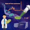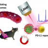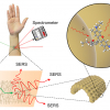
A team of researchers has demonstrated a new type of imaging system based on Raman spectroscopy that reveals the chemical composition of living tissue for medical diagnostics and cellular studies.
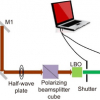
The new technique works by “coding” individual photons from a pulsing laser with a megahertz radio frequency and then collecting those photons with a detector after they have interacted with tissue. The system was demonstrated in human breast cancer detection. Ordinarily, the cancer tissue samples would have to be processed for histological examination, which could take up to a week. The new technology yields results in about two seconds.
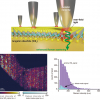
“We converted Raman spectroscopy, which is generally used to study molecules in solutions or fixed tissues, to an in vivo imaging platform that is able to monitor how a living cell executes its functions in real time. It’s a proof of concept. The approach allows us to get a spectrum of individual molecules, revealing the chemical composition of the tissues”, said Ji-Xin Cheng, a professor at Purdue University.
The results of their study are reported in a paper in Science Advances.
Their work is continuing, with future research exploring development of an “imaging pen” to quickly analyse surfaces for traces of explosives for homeland security applications and to monitor human tissue for disease and infection and to help surgeons determine whether any diseased tissue remains after cancer surgery, Cheng said.


