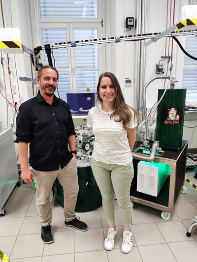
Researchers determine the structure and dynamics of proteins using nuclear magnetic resonance (NMR) spectroscopy. Until now, however, much higher concentrations were necessary for in vitro measurements of the biomolecules in solution than are found in our body’s cells. An NMR method enhanced by a very powerful amplifier, in combination with molecular dynamics simulation, now enables their detection and accurate characterisation at physiological concentrations. Dennis Kurzbach, a chemist at the University of Vienna and colleagues have demonstrated this new method with the example of a protein that influences cell proliferation and thus also potential tumour growth.
Currently, NMR spectroscopy is the only method that allows a complete description of the atomic structure of biomacromolecules in their native solution state. However, due to the inherently low sensitivity of the method, the samples must contain many more molecules per volume than physiologically common. To overcome this discrepancy, hyperpolarisation [more precisely by dissolution dynamic nuclear polarisation (DDNP)] can be used to achieve a 1000 × signal amplification in NMR measurements.
The researchers were able to measure biomolecules at concentrations as low as 1 µmole L–1 (one-millionth of the usual concentration levels). This concentration approaches that of our cells. This is important because proteins can react to unnaturally high concentrations. They no longer do what they are supposed to do and suddenly behave differently.
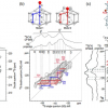
Besides, a dissolution DDNP measurement typically provides one-dimensional spectra, which limits the obtained information. To describe proteins comprehensively under natural concentration conditions, the researchers employed molecular dynamics simulations: “Such, we were also able to extrapolate the fingerprint we obtained of our molecule via NMR to its ‘entire body’, i.e. its multidimensional structure”, says Kurzbach.
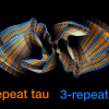
The value of this methodological advance is demonstrated using the ubiquitous transcription factor MAX. This protein can self-associate with various other proteins (i.e. protein dimerisation). For example, MYC-MAX dimers have a great influence on the DNA copying processes in the cell. With the new methods, MAX has been shown to adopt an undocumented conformation when concentrations approach physiological levels. “The folding spectrum of MAX is of crucial importance for working together with MYC and thus for the proliferation of healthy as well as diseased cells in the body”, said Dennis Kurzbach.
The new method can help to better understand the process of cell proliferation to tumour growth and thus elucidate basic mechanisms for cancer development. This is just one of many potential fields of application for the new method—after all, thousands of proteins in our cells perform a wide variety of tasks, including digestion and regulation of DNA and RNA.


