Imaging News
Remote simultaneous 3D and spectral imaging will provide direct identification of surface rust and corrosion on structures including bridges and pylons.
Terahertz imaging technology has the potential to help conservationists and academics better understand the history behind cultural artefacts.
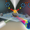
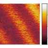
The National Physical Laboratory (NPL) in the UK has used tip-enhanced Raman spectroscopy to map catalytic reactions at the nanoscale for the first time.
A Lawrence Livermore engineer has been awarded $570,000 through the US Department of Energy SunShot initiative to explore spectroscopic technology as a means of detecting moisture build-up in solar cells.
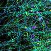
Research from Rice University, University of California, Los Angeles (UCLA) and Kansas State University in the USA has used super-resolution microscopy and fluorescence correlation spectroscopy to characterize such nanoscale spaces in chromatography media.
Renishaw recently broke ground on its new 133,000 ft2 office and warehouse facility in West Dundee, IL, USA, about 40 miles from Chicago. The two-storey facility will be located at the Oakview Business Park and will be the company’s North American headquarters. The new building, planned to be ready in June 2016, will also include space for product development, testing, warehousing and distribution. The facility will consolidate the company’s two existing sites and can also accommodate future expansion.
SPECIM, Spectral Imaging Oy Ltd, has received 5.3 M€ of financing to ensure its growth. The financing package consists of venture capital financing by Teknoventure Oy of Oulu, Finland, and R&D financing by Tekes, the Finnish Funding Agency for Innovation. The company, which was founded in 1995 by three former researchers of VTT Technical Research Centre of Finland Ltd, is known for its hyperspectral technology. The spectral imaging camera products are widely used in various industrial, environmental and defence applications.
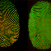
Watch the imprint of a tyre track in soft mud, and it will slowly blur, the ridges of the pattern gradually flowing into the valleys. Using imaging mass spectrometry, researchers at the National Institute of Standards and Technology (NIST) have tested the theory that a similar effect could be used to give forensic scientists something they have long wished for: a way to date fingerprints.
Raman microscopy is being used alongside high-resolution X-ray diffraction to unpick the reasons for crystallographic defects in SiC bulk crystal and epitaxial film, which limit the commercialisation of SiC devices.
Renishaw’s Spectroscopy Products Division (SPD) has moved to the new Renishaw Innovation Centre located at the company’s New Mills headquarters in Wotton-under-Edge, Gloucestershire, UK. This is a brand new building providing an additional 153,000 ft2 of space for Renishaw adjacent to the company’s iconic HQ, which is a converted 19th century woollen mill. The RIC was opened on 7 July 2015 by HRH The Princess Royal. SPDs new facilities also encompass a second, fully refurbished building, to house the Division’s production and supply chain teams.
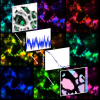
Combining fluorescence spectroscopy and “stochastic optical reconstruction microscopy” enables the imaging of single molecules with unprecedented spectral and spatial resolution, thus leading to the first “true-colour” super-resolution microscope.
A microscope being developed at the US Department of Energy’s Oak Ridge National Laboratory will allow scientists studying biological and synthetic materials to simultaneously observe chemical and physical properties on and beneath the surface.
Daylight Solutions, Inc., has received two new awards for their Spero™ microscope. Spero was awarded the 6th Annual Microscopy Today Innovation Award and was also named a finalist in the 2015 R&D 100 Awards taking place later this year.
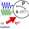
A new microscope based on ultrafast pump–probe spectroscopy and scanning near-field optical microscopy offers the potential for studying dyanmic processes on the nm scale.
Thanks to MALDI-MS imaging, scientists have discovered how earthworms can digest plant material, such as fallen leaves, that would defeat most other herbivores.
Diseases like Alzheimer’s are caused when proteins aggregate and clump together. Using atomic force microscopy and infrared spectroscopy, EPFL scientists have successfully distinguished between the disease-causing aggregation forms of proteins. The finding can help change pharmaceutical treatment of neurodegenerative diseases.
UCLA, USA, are combining Raman microscopy with scanning electron microscopy (SEM) to study archaeological textiles and fibres.
Synchrotron X-ray imaging of rocks is heping to save papers of the past.
Nano-Spectroscopy and Bio-Imaging is a free-to-attend conference in October 2015 held in Coventry, UK.
As part of around €24.5 million in funding for the next three years, the Deutsche Forschungsgemeinschaft (DFG, German Research Foundation) will establish one new Clinical Research Unit and nine new Research Units. As all DFG Research Units, the new units will collaborate interdisciplinarly and span multiple locations.
Among the new Research Units are three that may be of interest to readers (in alphabetical order by Host University).

