
 with jury members Katarzyna Marzec (second from right), Bo Peng (left) and WITec Managing Director Joachim Koenen (right).")
The Confocal Raman Imaging Symposium took place for the fifteenth time in Ulm, Germany, hosted by WITec who invited researchers, engineers and scientists to discuss the latest developments in confocal Raman microscopy from 24 to 26 September 2018.
With over 100 participants from many countries, attendance at the international conference was strong again this year. Well-known speakers from academia and industry informed the audience about research results in their respective fields. In addition, the participants were able to present their work through posters and discuss it with each other. The programme was supplemented by demonstrations of the latest confocal Raman microscopes. The evening lecture was given by the science comedian and physicist Vince Ebert, who entertained the international audience with precise and observant humour.
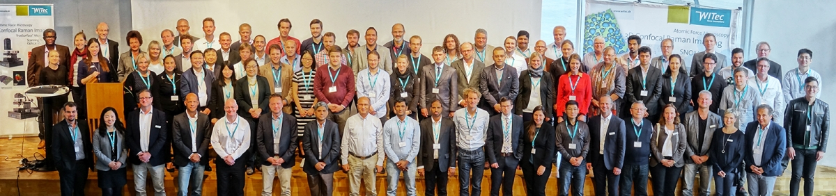
Group picture of the Symposium participants 2018.
At the beginning of the Symposium, Sebastian Schlücker (University Duisburg-Essen, Germany) gave a lecture on the theoretical basics of Raman spectroscopy and provided insights into special Raman techniques that increase the resolution and amplification of the Raman signal. Schlücker then let the audience test their knowledge with an interactive quiz.
Olaf Hollricher (WITec GmbH, Ulm, Germany) described the technical possibilities of Raman microscopes and explained how results can be improved with suitable instrument configurations.
Katarzyna Marzec (Jagiellonian University, Krakow, Poland) concluded the scientific presentations on the first day with a presentation of her work on correlative Raman microscopy in biomedical research. Marzec and colleagues use confocal Raman microscopy in combination with atomic force, near-field or fluorescence microscopy. They analyse biological tissues and cells to better understand and treat diseases such as arteriosclerosis, cancer and malaria.
The second day of the symposium started with a series of lectures on nanotechnology and low-dimensional materials. The lectures by Dirk Guldi (University of Erlangen, Germany) and Bo Peng (University of Electronic Science and Technology, Chengdu, China) dealt with research work that aims to improve the material properties of graphene, tungsten disulphide and molybdenum disulphide. These conductive nanomaterials are to be used in electronic devices in the future.
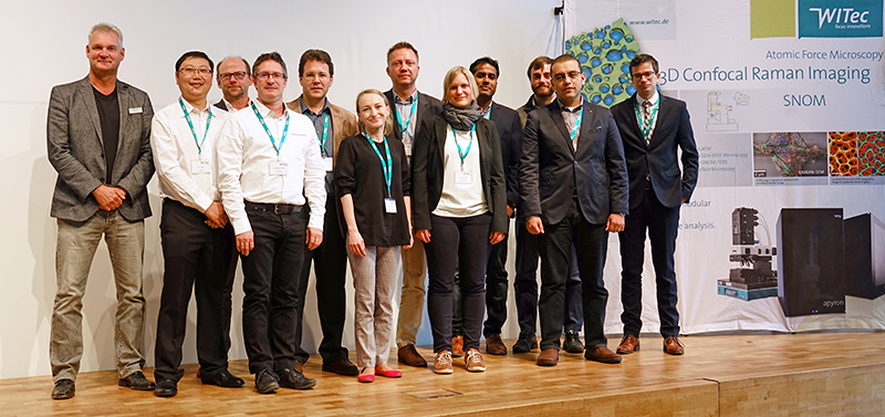
The speakers of the Confocal Raman Imaging Symposium 2018: (from left to right): Olaf Hollricher, Bo Peng, Dieter Baurecht, Guillaume Wille, Dominic Papineau, Katarzyna Marzec, Martin Maiwald, Eva Brauchle, Vinayam B. Parambath, Isaac Pence, Hesham K. Yosef, Christian Timma. Not pictured: Sebastian Schluecker, Vince Ebert, Dirk Guldi, Keith Gordon, Jagjit Nanda.
To begin the geoscientific lecture series, Guillaume Wille (BRGM Orleans, France) presented his investigations of polymorphic structures and asbestos using correlative Raman Imaging and Scanning Electron (RISE) microscopy. He combines the electron microscopy findings on sample morphology and nanostructure with the information on chemical mineral composition obtained by Raman microscopy. His investigations on asbestos allow health risks to be better identified and prevented.
Dominic Papineau (London Center for Nanotechnology, UK) delivered an overview of his comprehensive research results on geological samples. He is interested in the detection of organic compounds in rock in order to date the primeval occurrence of microbial life more precisely. Using Raman microscopy, Papineau and colleagues have recently been able to identify one of the oldest microfossils ever found.
Keith Gordon (University of Otago, Dunedin, New Zealand) detailed his chemical analyses of a wide range of different samples, including Maori archaeological fibres and hypomineralised teeth.
The materials science lecture series featured Martin Maiwald (Ferdinand-Braun-Institute, Berlin, Germany), who reported on SERDS, a method for removing interfering background and fluorescence signals from a Raman spectrum by means of two slightly different excitation wavelengths, which helps achieve better results.
Jagjit Nanda (Oak Ridge National Laboratory, USA) has been researching batteries for many years and was able to present extensive Raman analyses on the charge state of lithium-ion batteries.
Christian Timma (thyssenkrupp Steel Europe AG, Duisburg, Germany) vividly reported on the establishment of confocal Raman microscopy for quality control in steel production in his company.
In the life sciences lecture series, Isaac Pence (Imperial College London, UK) explained his research results on tissue-engineered cartilage. Pence and his colleagues hope to obtain an accurate picture of the chemical and molecular components of the tissue in order to come as close as possible to the properties of natural tissue. Pence also used Raman microscopy to analyse differences in the lipid composition of the myelin layer between normal nerve tissue and the tissue of mice with multiple sclerosis. He also presented the SPARTA method, which allows the analysis of small nanoparticles in solution by Raman spectroscopy without staining.
Dieter Baurecht (University of Vienna, Austria) talked about the broad spectrum of questions that are brought to his laboratory from different disciplines that he tries to solve by using confocal Raman microscopy and AFM.
This was followed by short presentations selected by a jury from the submitted summaries of the research work of the Symposium participants. Hesham K. Yosef (Ruhr University Bochum, Germany) gave a lecture on pharmacokinetic investigations of lung and breast cancer cells. He uses correlative Raman fluorescence microscopy to analyse the uptake and conversion of the cancer drug neratinib in cell cultures and was able to show that receptors that are important for cell growth are degraded by its introduction. Eva Brauchle (University of Tübingen, Germany) presented her work on Parkinson's disease. Using Raman imaging, she investigates altered protein structures and the formation of aggregates in the neuronal tissue of rats. In the future, her research might help to obtain information about the course of the disease at an early stage. Vinayam B. Parambath (HIU, Ulm, Germany) researches the charging and discharging mechanisms of metal-sulphur batteries in order to make them more efficient in the future.
In the evening, the traditional conference dinner took place in the Ratskeller in Ulm. The Poster Award was also presented there. This year, the jury chose Dieter Fischer’s (Leibniz Institute of Polymer Research, Dresden, Germany) poster on microplastic particle analysis.
The abstract book can be downloaded from https://www.raman-symposium.com/assets/Uploads/WITec-AbstractBook-15ConfocalRamanImagingSymposium.pdf.
